


#17 Making Visible Invisibility, Animals Looking After Their Homes and The Genetics Linked To Hearts...
Making invisible blood vessels visible, animals have evolved to not overexploit and genetic variants involved in congenital heart disease.
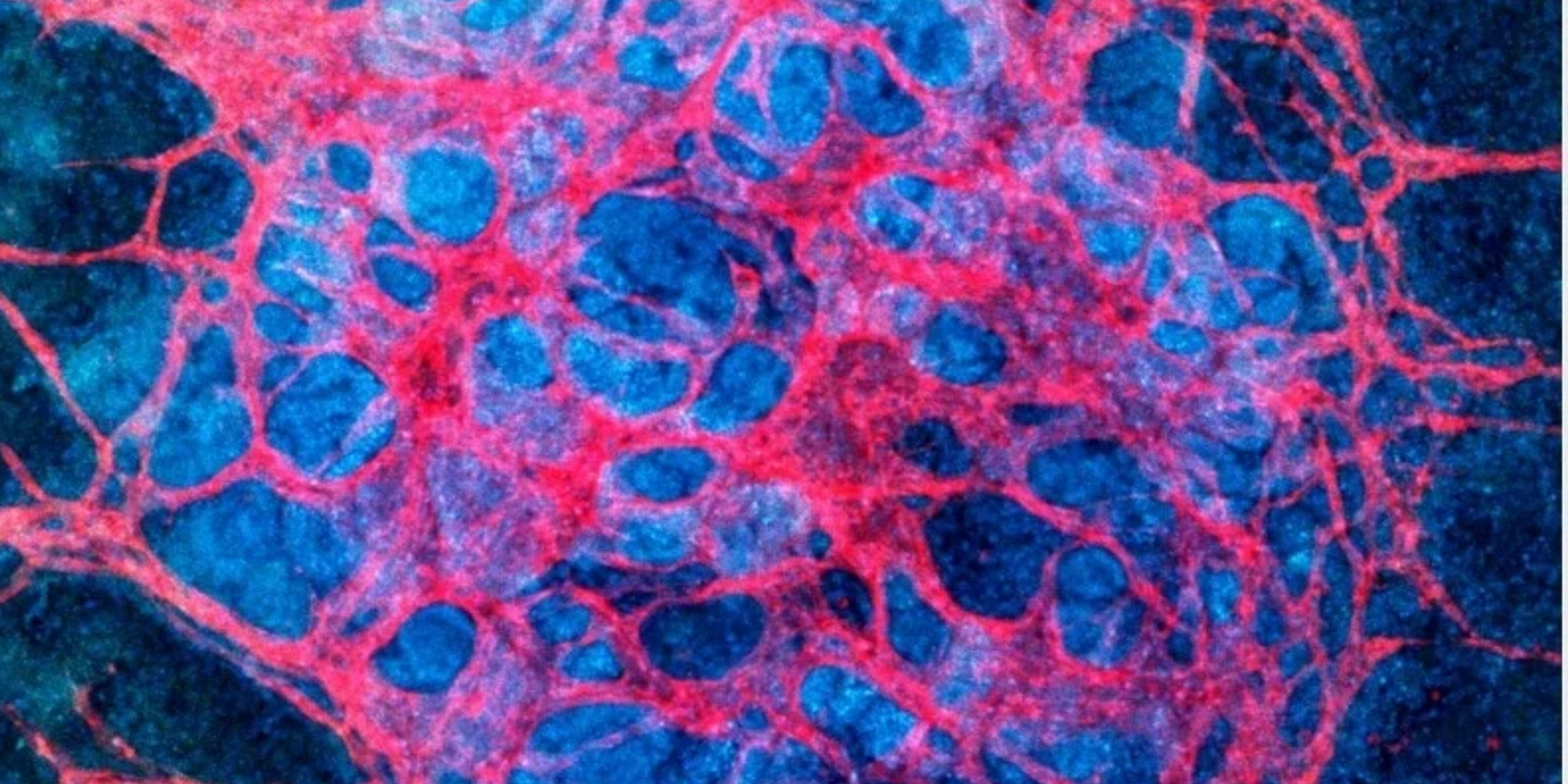
🩸Visible Invisibility
Making Invisible Blood Vessels Visible…

Whilst blood vessels cover a large surface area they are stilly relatively tiny, with the smallest blood vessels covering only five micrometres. That size being roughly 1/3 the size of a single strand of human hair.
Researchers from Johns Hopkins Medicine have tested and developed a new imaging approach that will supposedly accelerate image based research in the lab. This allows the capturing of blood vessel images at different spatial scales.
The method has been dubbed “VascuViz” and includes a polymer mixture to fill blood vessels, making them visible in multiple imaging techniques. This approach enables the researchers to visualise the structure of a tissue’s vasculature, with other information this can help the scientists clarify the complex role of blood flow in health and disease. These combined issues should not only enhance the study of the diseases, such as those involved with blood flow abnormalities - cancers and strokes, but also help to advance our understanding of the structure and function of tissues throughout the body.
Senior Author, Arvind Pathak said that “Usually, if you want to gather data on blood vessels in a given tissue and combine it with all of its surrounding context like the structure and the types of cells growing there, you have to relabel the tissue several times, acquire multiple images and piece together the complementary information,”. He went on to say how this can be an expensive and time consuming process that can risk destroying the architecture of the tissue and so precluding the ability to use the combined information in novel ways.
Previously, researchers use multiple different methods such as magnetic resonance imaging (MRI), computed tomography (CT) and microscopy to study the role of vessels in the lab. These images can help in understanding the dynamics and ways in which the tissue responds to disease, but often make the vessel unusable for multiple methods of research or imaging. This is due to the fact that one method of imaging method can often make it unusable for other imaging methods. This limits the amount of data researchers can gather from a single sample.
The new VascuViz method overcomes this issue and integrates the different types of data available. VascuViz makes the largest structures as well as the smallest microvasculature visible to a variety of imaging tools. This allows researchers to develop a multi-layered understanding of blood vessels and related tissue components. Complex systems in biology such as the circulatory system can now be better understood through the creation of computerised visualisations. This new method can now act as a hallmark in the growing field of image-based vascular systems biology.
The studies lead author; Akanksha Bhargava (Johns Hopkins University School of Medicine) went on to say that “Now, rather than using an approximation, we can more precisely estimate features like blood flow in actual blood vessels and combine it with complementary information, such as cell density,” “To do this, VascuViz-based measurements are entered into computer simulations of blood flow” such as the cancer models Bhargava studies.
To create VascuViz, Bhargava tested several combinations of existing imaging agents and their suitability for different imaging methods. With research, time and adaptations she found that a CT contrast agent named BriteVu as well as a fluorescently labelled MRI contrast agent named Galbumin-Rhodamine could create a compound that would make both the macro and microvasculature visible when imaging through all methods and without interference or damage to the tissue samples.
Following successful lab tests and with the compound effectively working in test tubes, the researchers tested the compound on a variety of mouse tissues, perfusing it through the vascular system of breast cancer models, leg muscles, brain and kidney tissues. Resulting images of the tissues acquired through the various methods previously mentioned, were combined to create stunning 3D models and visualisations of the vasculature and associated components comprising these disease models and organ systems. Due to the affordability and commercially available components the team hopes that it will be globally adopted by scientists to help shed new light on different diseases involving the vasculature.
🛖Animals Look After Their Homes
Animals have evolved to avoid overexploitation…

The equilibrium between predator and prey has been a relationship studied for thousands of years. With Charles Darwin publishing his revolutionary theory of evolution “On the Origin of Species” many questions regarding this relationship were raised. Including the question “Why do predators not evolve, become so aggressive that they eat all the prey and go extinct?”.
The American ecologist Lawrence Slobodkin proposed the idea of prudent predation in 1960, where predators are able to avoid extinguishing their own prey. Biologists argued that prudent predation would require evolution to act on groups rather than single individuals of a species and so “group selection” was unlikely to occur. Whilst modern evolutionary theory has moved beyond this, with commonly held scepticism, the theory of prudent predation is still a held belief by some scientists.
In a recent study published in Ecology Letters, through the use of a complex predator-prey model, the research team showed how this predator-prey equilibrium could have occurred.
Prudent predation is defined as a predator species evolving to not consume as much and as aggressively as its own physical limits permit. In doing so restraining themselves for the benefit of other members of their own species and so future generations - this is not done so knowingly.
Predators are often prudent in their natural habitat, however less so when moved to foreign and unnatural environments. This is demonstrated by the Indo-pacific lionfish whose population numbers have drastically increased in and around the Gulf of Mexico and the eastern Mediterranean Sea. Lionfish typically feed on smaller fish and shellfish that live in reefs. They are renowned, ferocious predators to the point where ecologists became worried as to whether other fish species would survive their presence, especially in the Gulf of Mexico. Rather, the population dynamics changed, much to the surprise of the ecologists.
Lionfish populations suddenly fell, whilst their native competitors remained. It appeared that due to lionfish overexploiting prey, they are not such strong competitors after all. These dwindling lionfish populations are therefore experiencing evolutionary pressure to feed less ferociously, so that they can occupy reefs for longer periods of time and have more opportunities to spread to other locations and coral reef sites. Eventually it is expected that they will adapt to their new habitat becoming prudent predators.
🖤Genetics Involved in Heart Issues
Genetic variants involved in congenital heart disease…

Congenital heart disease means a heart condition or defect that develops in the womb, before the baby is born. There are numerous examples of these diseases with issues varying from unformed valves to holes between chambers. Nearly 1% of all children are born with congenital heart diseases. It remains that for most children, the precise cause of these frightening defects are unknown.
The causes of these conditions appears to be abnormal or variants of genes involved with the formation of the structure and functioning of the heart. However, there is still much to learn in terms of exactly which genes contribute to congenital heart disease and how they interact with each other.
The Gladstone Institute have developed a novel method for identifying genetic variants that are likely to play important roles in congenital heart disease. This opens up opportunities to accelerate research into this serious condition. The new strategy combines techniques from genetics, computational biology, stem cell biology and proteomics and could be applied to study numerous other diseases with complex genetic causes.
"Previous methods have generated long lists of variants detected in patients, but many actually turned out to be inconsequential, so a major challenge in the field has been identifying which variants are most important," says Srivastava, the Gladstone President and Senior Investigator. "Our approach pinpoints variants that are most likely to be involved in disease, allowing us to focus on those variants, deepen understanding of the underlying biology of the disease, and, we hope, move more rapidly toward new treatments."
Rather than looking at variants in isolation the novel strategy considered the interactions between proteins to focus in on which variant may be the cause of the disease. The proteins GATA4 and TBX5 were already known to be required for healthy heart formation and collaborates with a network of additional proteins to help the heart grow. Mutations in these other proteins could contribute to heart malformation.
To identify these potentially culprit genes, the researchers carefully mapped out the entire network of interactions between the GATA4 and TBX5 proteins using precursor heart cells grown from human induced pluripotent stem cells. Next they cross-referenced this 273 protein network with DNA sequencing data from over 3,000 children with congenital heart disease. Several dozen variants in the children’s sequencing data matched those of specific proteins in the GATA4-TBX5 network and so pinpointing those as potential heart disease causing candidates.
Determining which candidate variants identified actually contribute to heart disease could take years of research. Instead Maureen Pittman, UCSF graduate student, developed a computational tool that ranks the candidates according to their likelihood of congenital heart disease contribution. Pittman said "But many had never before been linked to heart development, including a protein called GLYR1, which is involved in turning other genes on and off." Additional experiments in cells and mice indicated that GLYR1 indeed plays a key role in the formation of the heart, with a patient variant of GLYR1 disrupting heart development by hampering its interactions with GATA4.
Thanks to advancements in the area and in surgery specifically, millions of children with heart defects survive to adulthood. Many however still face lifelong problems with an increased risk of heart failure. The researchers believe that the power of their new method lies in its promise to help illuminate how combinations of variants, rather than single variants on their own, work to cause congenital heart disease.
Weekly Topics
🏞️ Environmental
Scientist uses trackers to track funnel web spiders
Polar bear inbreeding and bird divorces - climate change
Ancient fish lizard - reconstructed…
🦭Marine
One in two million - blue lobster
🐼 Conservation
15% of global coastal regions remain intact
Large mammals can help mitigate climate change
Fresh banana leaves has harmed Indigenous people
🦠 Disease and Illness
Lymphatic disorder may cause stillbirth or disease in affected children
Mitochondrial diseases are transmitted from mother to child
Infectious disease - “Many Voices, One Health”
😷 COVID
COVID19 and the life sciences - What have we learned…
How does COVID cause a loss of smell
Omicrons molecular structure could explain its global takeover
🧪 Biochemistry
New astrobiology predicts life…
Scientists engineer bacteria to cope in challenging environments
Pioneer simulations focus on HIV-1 Virus
🔬 Evolution
The mechanisms capturing the mechanism underlying evolvability
Gradual evolution is back - Darwinian Theory
Rapid evolution fuels transcriptional plasticity in fish species
🧬 Genetics
New tool reveals function of enigmatic gene sequences
Exploring genetics and atopic dermatitis
Simple reverse genetics method to generate recombinant corona viruses
📷 Weekly Camera Roll
Click on the text below to keep reading…


Reference List
Content may be adapted and edited for style and length.
🩸Visible Invisibility
Vascular News (1st March 2022) https://vascularnews.com/making-the-invisible-visible/
Bhargava, A., Monteagudo, B., Kushwaha, P. et al. VascuViz: a multimodality and multiscale imaging and visualization pipeline for vascular systems biology. Nat Methods 19, 242–254 (2022). https://doi.org/10.1038/s41592-021-01363-5
🛖Animals Look After Their Homes
The Conversation - Axel G. Rossberg (2022) Axel G. Rossberg Reader in Theoretical Ecology, Queen Mary University of London. Available from https://theconversation.com/animals-have-evolved-to-avoid-overexploiting-their-resources-can-humans-do-the-same-176092
🖤Genetics Involved in Heart Issues
Gonzalez-Teran, B., et al. (2022) Transcription factor protein interactomes reveal genetic determinants in heart disease. Cell. doi.org/10.1016/j.cell.2022.01.021.